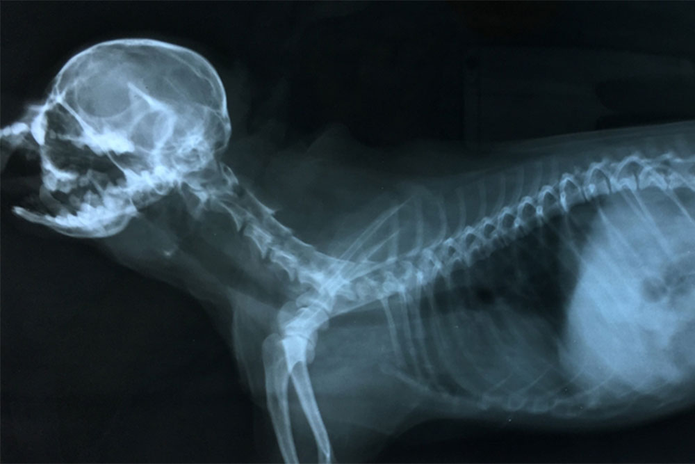
If your pet is sick or injured, your veterinarian may order a digital X-ray, an ultrasound, or a computed tomography (CT) scan to make an accurate diagnosis. These advanced medical imaging techniques are a noninvasive way your veterinarian can observe your pet’s internal structures to determine a health problem’s cause. Neighborhood Veterinary Centers describes these techniques to help you understand when these imaging procedures are indicated, and how they work.
Digital X-rays for pets
The X-ray (i.e., radiograph) is the most common imaging technique veterinarians use to detect bony abnormalities, visualize organs, distinguish foreign bodies in the intestinal tract, assess dental disease, and detect chest or abdominal fluid accumulation. Radiography generates a black and white, two-dimensional image by passing radiation through a particular body area. Traditional X-rays record the image on special X-ray film that senses the radiation amount passing through the structure and reaching the film. Dense tissues, such as bone, appear white on the film, and less dense structures, such as air-filled lungs, appear black. Digital X-rays are captured on a digital recording device, and displayed on a computer screen. Benefits over traditional imaging techniques include:
- Faster diagnosis — Digital technology presents the images immediately, and because they do not have to wait for film development, your veterinarian can diagnose your pet’s condition more quickly.
- Convenience — Unlike traditional radiograph film images, digital X-ray images can be stored conveniently in your pet’s file.
- Improved quality — Your veterinarian can manipulate digital X-ray images, improving the quality to get a clear view.
- Easier measurement calculation — Using digital technology, your veterinarian can take measurements and make calculations while viewing the images.
- Better resolution — Digital images provide clearer resolution, allowing your veterinarian to observe more detail on your pet’s X-ray than on traditional X-ray images.
- Safer — Digital radiography systems expose your pet to less radiation than traditional X-ray systems.
Many X-ray views can be taken without sedation, but if your pet is anxious or painful, or if the views require extensive manipulation, your veterinarian may administer a sedative or anesthesia.
Ultrasound for pets
Diagnostic ultrasound uses piezoelectric crystals from a probe (i.e., transducer) to send sound waves through your pet’s body. When the sound waves hit boundaries, such as between fluid and soft tissue, or between soft tissue and bone, they echo back to the transducer, and generate electrical signals that are sent to the ultrasound scanner. The unit calculates the distance from the transducer to the tissue boundary, generating a two-dimensional image of your pet’s internal structures. Your pet may need an ultrasound for reasons such as:
- Heart conditions — If your pet has a heart murmur or heart arrhythmia, your veterinarian may recommend a heart ultrasound (i.e., echocardiogram) to determine the abnormality’s cause.
- Organ assessment — An abnormal blood or urine test may indicate your pet has a condition affecting a particular organ. Your veterinarian may perform an abdominal ultrasound to visualize your pet’s organs including the liver, kidneys, spleen, lymph nodes, and urinary bladder.
- Foreign body — If your pet potentially ingested a foreign body, your veterinarian may perform an ultrasound to assess the gastrointestinal tract to find the blockage.
- Eye injury — Your veterinarian can use ultrasound to assess your pet’s eye if disease or injury prevents assessment through an ophthalmoscope, or if the structures surrounding the eye are involved.
- Soft tissue injury — Your veterinarian may perform an ultrasound to assess damage to your pet’s tendons and ligaments.
- Abdominal masses — If your veterinarian detects an abdominal mass when palpating your pet’s abdomen, they may recommend an ultrasound to observe the mass’s characteristics.
- Fetal assessment — Your veterinarian can use ultrasound to assess a fetus in your pregnant pet, determining viability and development stage.
- Emergency situations — In an emergency situation, your veterinarian may use ultrasound to make a quick determination whether your pet has internal hemorrhage or injuries.
- Sample collection — Your veterinarian can use ultrasound to guide sample collection when obtaining a needle aspirate or for some biopsies.
Before the ultrasound, your veterinary professional will shave your pet’s hair in the evaluation area so the probe can make better skin contact. Most pets tolerate an ultrasound examination well, but some require sedation.
Computed tomography for pets

Computed tomography (CT) is a computerized X-ray imaging technique in which a narrow X-ray beam is aimed at your pet, and quickly rotated around their body to generate cross-sectional images or slices. A CT scan provides more detailed information than conventional X-rays, and the slices can be stacked together to form a three-dimensional image, so your veterinarian can more easily identify basic structures and potential abnormalities. Veterinarians often order CT images in the following circumstances:
- Nasal cavity and sinus disease — Your veterinarian may use CT technology to assess the complex anatomy in your pet’s nasal cavity and sinuses.
- Spinal disorders — CT images, especially when using contrast dyes, are helpful in the diagnosis of spinal injuries that occur with conditions such as intervertebral disc disease.
- Lung abnormalities — Your veterinarian can use CT images to evaluate your pet’s lung tissue in great detail to detect issues such as lung tumors and pulmonary fibrosis.
- Joint disease — Your veterinarian can use CT technology to evaluate your pet’s joint in detail, to detect conditions such as elbow dysplasia, hip dysplasia, and osteochondritis dissecans.
- Tumor evaluation — Veterinarians frequently use CT imaging to plan surgical removal and radiation treatment for tumors.
To ensure your pet remains completely still when undergoing a CT scan, your veterinarian will administer anesthesia.
Advanced imaging techniques are helpful to ensure your pet receives an accurate diagnosis. If you believe your pet has a condition that may benefit from an advanced imaging procedure, contact our Neighborhood Veterinary Centers team so we can ensure they get the care they need.


Leave A Comment